
Brendan Leng Yong Ji
Overview
Gonioscopy is used to visualise the drainage angle of the anterior chamber. It is commonly utilised in the context of glaucoma to grade the anterior chamber angle and to diagnose angle closure. It is a vital skill for all ophthalmologists and good understanding is indispensable in ensuring prompt identification and diagnosis of angle closure.
Principles
The structures of the iridocorneal angle are not normally visible due to total internal reflection. Total internal reflection occurs when the angle of incidence of light passing from a medium of greater density to one of lesser density is larger than the critical angle of the interface between the two mediums. This results in the reflection of light back into the original denser medium. (1) The critical angle of the air-cornea interface is around 46 degrees (2), hence light from the iridocorneal angle reflects back into the anterior chamber, disallowing direct visualisation. Placing a lens minimises the refraction at the cornea, creating a new air-lens interface that allows visualisation of the iridocorneal angle by an observer.
Direct vs Indirect Gonioscopy Lens (3-5)
There are two major categories of lenses utilised in gonioscopy, direct and indirect lenses. Direct lenses allow direct visualisation of the iridocorneal angle and provide an erect view of the structures. Direct gonioscopy allows for a panoramic view of the angle, greater control over angle of visualisation and a direct view for surgical intervention. Placing lenses on both eyes simultaneously enables comparison between both eyes which can help identify angle recession. The major drawback to direct gonioscopy is the requirement for specialty equipment and the need for the patient to remain supine. Due to its inconvenience, direct gonioscopy is usually done in theatre. Direct gonio lenses include Koeppe, Barkan, Shaffer, Thorpe and Swan Jacob.
Indirect gonioscopy lenses are more common in clinical practice due to their relative ease of use. The image seen by the observer will be inverted as the image is reflected off a mirror. However, advantages of indirect lenses include convenience, greater optics and clarity and the possibility of dynamic gonioscopy. Patients are not required to lie supine with indirect lenses and less instrumentation is required. Furthermore, dynamic gonioscopy can help differentiate between fixed and non-fixed causes of angle closure. Limitations of indirect lenses include only providing a segmental view, only being able to examine one eye at a time and inadvertent indentation may open the angle giving a false evaluation of the angle. Examples of indirect lenses include Posner, Sussman, Zeiss and Goldman.
Methodology (3-5)
Direct Gonioscopy
1. Ensure a binocular microscope or a slit-pen light is available along with a direct goniolens
2. The procedure is commonly conducted with the patient under anaesthesia
3. Place the patient in a supine position
4. Introduce saline or viscoelastic on the eye to act as a coupling agent
5. Position the lens onto the eye
6. Adjust the microscope and light source to examine the entire angle
Indirect Gonioscopy
1. Ensure the room is dimmed and that the patient is positioned comfortably
2. Apply topical anaesthetic to both eyes
3. If using a lens with a surface larger than the cornea (i.e. Goldmann) use a gonioscopic coupling gel
4. If using a lens with a smaller surface than the cornea (i.e. Zeiss or Sussman) the patient’s tear film can function as the coupling agent, though artificial tears can also be applied
5. Use the shortest slit beam possible, high magnification and off-centre the beam by 30-35º
6. Ask the patient to look up, place the lens onto the eye then ask the patient to look straight
7. Stop moving the lens once the iris is in view
8. Identify all angle structures in all segments of the eye
9. Repeat steps for other eye
Dynamic Gonioscopy
1. A Posner, Sussman or Zeiss lens is required for dynamic gonioscopy
2. Procedure is similar as with indirect gonioscopy
3. Gentle pressure is placed on the cornea with the lens which forces aqueous into the angle
4. If the angle closure is fixed then the angle will remain the same
5. If the angle closure is non-fixed or appositional then the angle structures will become more visible
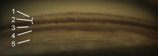
Structures of the Angle
Schwalbe’s line
Schwalbe’s line is the peripheral termination of descemet’s membrane and appears as an opaque line. A prominent and anterior schwalbe’s line is called a posterior embryotoxon and can be a normal variant or associated with Axenfield-Rieger syndrome (7). If there is excessive pigmentation at or anterior to shwalbe’s line this is termed Sampolesi line and is associated with pseudoexfoliation syndrome or pigment dispersion syndrome (5).
Trabecular Meshwork
The trabecular meshwork (TM) consists of an anterior non-pigmented and posterior pigmented part. The non-pigmented TM lies posterior to schwalbe’s line and pigmented TM lies anterior to the scleral spur. The pigmented TM gains its pigmentation throughout life but can be secondary to conditions such as uveitis, pigment dispersion syndrome and pseudoexfoliation syndrome (8).
Schlemm’s Canal
Schlemm’s canal is not normally visible but can occasionally be seen posterior to the TM as a red line in non-pigmented eyes or in pathological conditions. Conditions such as Sturge-Weber syndrome or cavernous fistulas can result in blood regurgitating into schlemm’s canal, resulting in its red appearance (9).
Scleral Spur
The scleral spur is the site of attachment for ciliary muscles and is located posterior to the and anterior to the ciliary body. It appears white due to its high collagen content (5).
Ciliary Body
The anterior ciliary body can be seen as a dull pink, brown or grey band in eyes with deeper angles. A particularly deep angle or asymmetrically deep angles should highlight the possibility of angle recession or cyclodialysis (3).
Grading
There are many grading systems used to categorise the appearance of the iridocorneal angle. Some of the most commonly utilised in clinical practice include the Scheie, Shaffer, Shaffer-kanski, Spaeth, Becker and Van Herick system. The Shaffer-kanski system is based off the Shaffer system which grades the angle from 0-4 based on angularity. A grade 4 angle is wide open with no risk of angle closure whilst a grade 0 angle is closed with no visible structures. An in depth overview of the different grading systems can be found in the Atlas of Clinical Gonioscopy by Wallace L M Alward (10).
| Grade Angle (°) | Angularity | Structures Visible | Risk of angle closure |
| 0 | 0 | No structures visible | Closed angle |
| 1 | 10 | Schwalbe’s line, possibly anterior trabecular meshwork visible | Closure possible |
| 2 | 20 | Schwalbe’s line and trabecular meshwork visible | Closure unlikely |
| 3 | 20-35 | Schwalbe’s line, trabecular meshwork and scleral spur visible | Closure impossible |
| 4 | 35-45 | All structures visible | Closure impossible |
Conclusion
Gonioscopy is an indispensable tool in the diagnosis of angle-closure glaucoma. Good understanding of the different tools available and the structures visualised is vital in accurate identification and diagnosis. Grading systems are available that can assist with the stratification of risk in relation to clinical findings.
References
1) Elkington AR, Frank HJ, Greaney MJ. Clinical Optics. 3rd ed. Philadelphia, PA: Blackwell Science; 1999.
2) Sinha A, Mohan S, Gupta V, Sihota R. Gonioscopy. Delhi Ophthalmological Society Times. 2005 Mar;10:322–8.
3) Ahmad SS, Queen Elizabeth Hospital, Kota Kinabalu, Malaysia. Gonioscopy—A Primer. US Ophthalmic Rev [Internet]. 2017;10(01):42. Available from: https://dx.doi.org/10.17925/usor.2017.10.01.42
4) Unterlauft JD. Gonioscopy – how, why, what for? Klin Monbl Augenheilkd [Internet]. 2017;234(8):996–1002. Available from: https://dx.doi.org/10.1055/s-0042-120280
5) Greenwood M, Gupta S, Aref A, Siegfried C, Tripathy K, Seibold L. Gonioscopy – EyeWiki [Internet]. American Academy of Ophthalmology. 2021 [cited 2022 Feb 3]. Available from: https://eyewiki.aao.org/Gonioscopy
6) Supakontanasan W, Thunwiriya P, Suwan Y, Nilphatanakorn S, Arunmongkol S, Teekhasaenee C. Effect of visibility of the ciliary body processes on ocular biometric parameters in patients with primary angle closure. Jpn J Ophthalmol [Internet]. 2019;63(6):467–73. Available from: https://dx.doi.org/10.1007/s10384-019-00686-3
7) Rennie CA, Chowdhury S, Khan J, Rajan F, Jordan K, Lamb RJ, et al. The prevalence and associated features of posterior embryotoxon in the general ophthalmic clinic. EYE [Internet]. 2005;19(4):396–9. Available from: https://dx.doi.org/10.1038/sj.eye.6701508
8) Shihadeh W, Ritch R. Reducing the release of pigment [Internet]. Glaucoma Today. 2005 [cited 2022 Feb 3]. Available from: https://glaucomatoday.com/articles/2005-july-aug/0705_06.html
9) Deaner J. September 2014 wills eye resident case series – diagnosis & discussion [Internet]. Reviewofophthalmology.com. 2014 [cited 2022 Feb 3]. Available from: https://www.reviewofophthalmology.com/article/september-2014-wills-eye-resident-ca se-series–diagnosis–discussion
10) Alward WLM. Color Atlas of Clinical Gonioscopy. Mosby-Year Book; 1993.
11) Bowling B. Kanski’s clinical ophthalmology: A systematic approach. 8th ed. London, England: W B Saunders; 2015