Abdullah A. Cheema1, Connor Qiu2, Mohamed Ahmed Roshdy Khalil3
1Royal Glamorgan Hospital, Llantrisant, Wales
2Royal Free London NHS Foundation Trust, London
3Glangwili Hospital, Carmarthen, Wales
Abstract
Phototoxic retinopathy may occur following cataract surgery. Optical coherence tomography (OCT) was performed in two patients with clinical and fluorescein angiographic phototoxic retinopathy subsequent to phacoemulsification cataract surgery. Both eyes demonstrated neuro-sensory detachment from underlying RPE in the affected retina, with lesser and varying degree of neural retinal thickening and cystoid change. OCT may be helpful in the diagnosis and follow up of this condition.
Introduction
Retinal phototoxicity induced by operating microscope illumination has been described subsequent to cataract surgery (1, 2, 3, 4). Fluorescein angiographic findings in phototoxic retinopathy have also been well documented in the literature (1, 4, 5). The purpose of this case series is to report the ocular coherence tomographic (OCT) findings with StratusTM OCT (Carl Zeiss Ophthalmic Systems, Inc. Dublin, CA) in post operative phototoxic retinopathy, and their correlation with clinical and fluorescein angiographic findings. We are not aware of any previous report of OCT findings in this context.
Case Reports
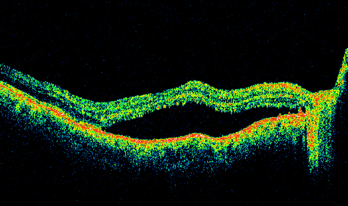
Case 1: A 75 years old healthy woman underwent routine phacoemulsification with lens implant for a dens nuclear sclerotic cataract in left eye. Post operatively the visual acuity was 6/18 at 10 weeks. Fundal examination showed an oval area 2 disc diameter in size showing yellowish changes in infero-temporal aspect of fovea. (Figure 1A) Fluorescein angiography revealed changes consistent with diagnosis of photo-toxic retinopathy, showing a corresponding but larger area of patchy hyperfuorescence and hypofluorescence in the early and mid phase. This was in an ovoid and semicircular configuration. Late phase angiogram showed punctate staining, and minimal leakage. (Figure 1B) OCT was carried out at 10 weeks post operatively. Fast macular thickness map revealed an oval area of thickening extending from inferior fovea to a 3mm diameter. The most affected sector was temporal inner macula, mean retinal thickness was 347µ compared to 147µ on the right (Figure 1C). A 6mm scan revealed dome shaped detachment of neurosensory retina, with a small degree of thickening of neurosensory retina (Figure 1D).
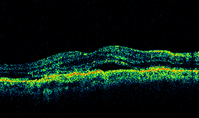
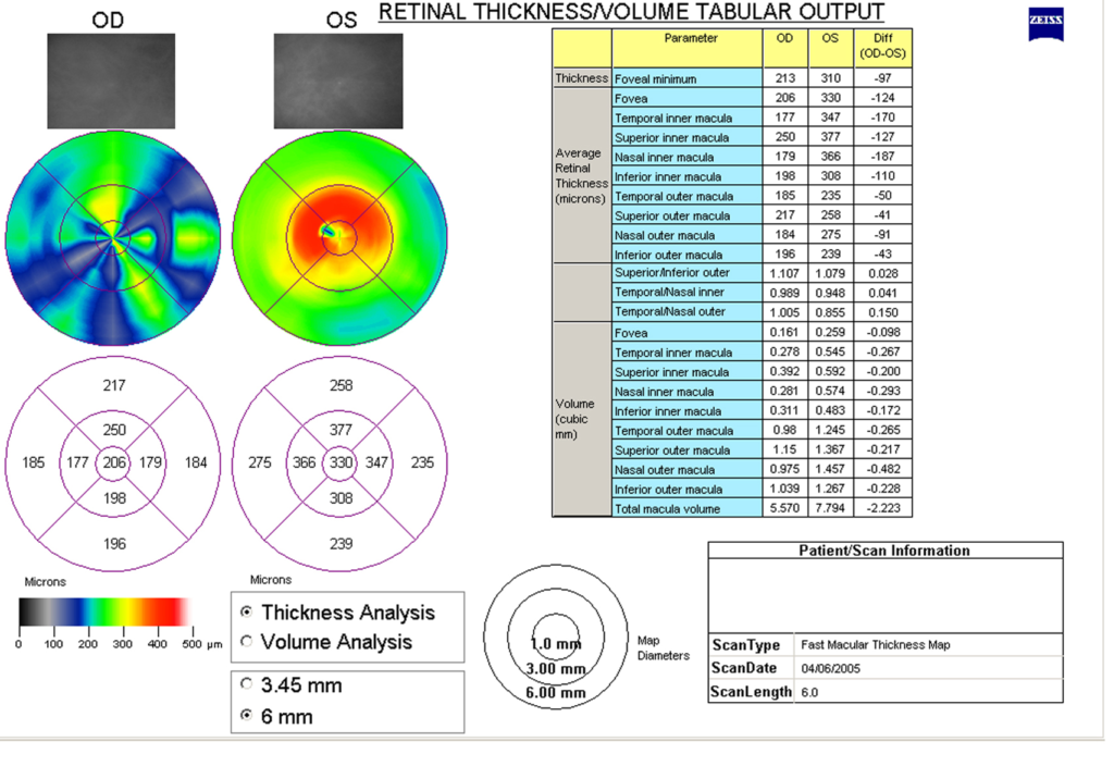
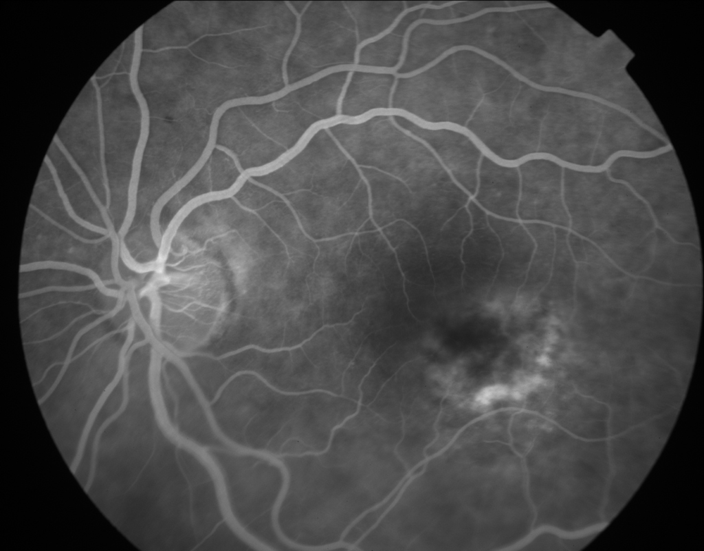
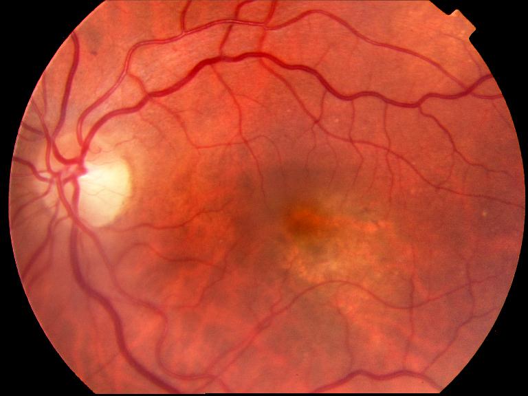
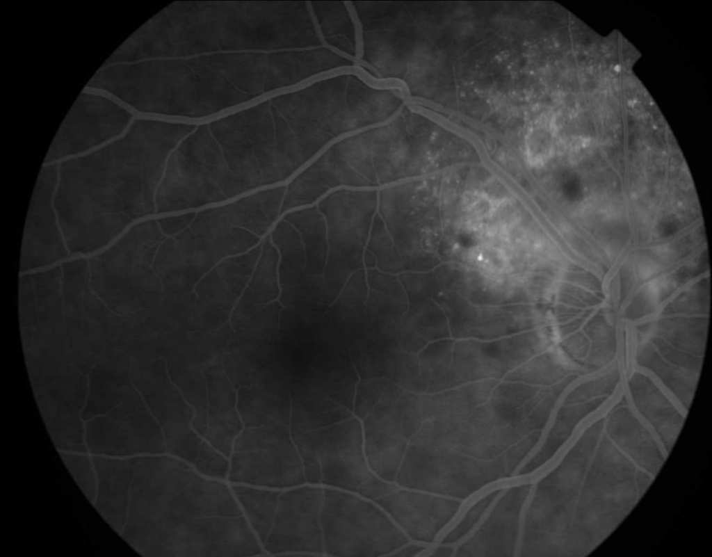
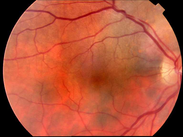
Case 2: An 82 year old man with nuclear sclerotic cataract underwent routine phacoemulsification and lens implant using temporal corneal approach in the right eye. Post operative visual acuity was 6/12 at four weeks. Fundal exam showed retinal elevation with yellowish pigmentary changes involving an oval shaped area nasal to the fovea along superior temporal arcade (Figure 2A). Fluorescein angiography revealed large area of mottled early hyperfluorescence, with hypofluorescent patches in the affected retina. Late phase angiogram showed punctate staining, but no leakage. (Figure 2B). A diagnosis of Phototoxic retinopathy was made.
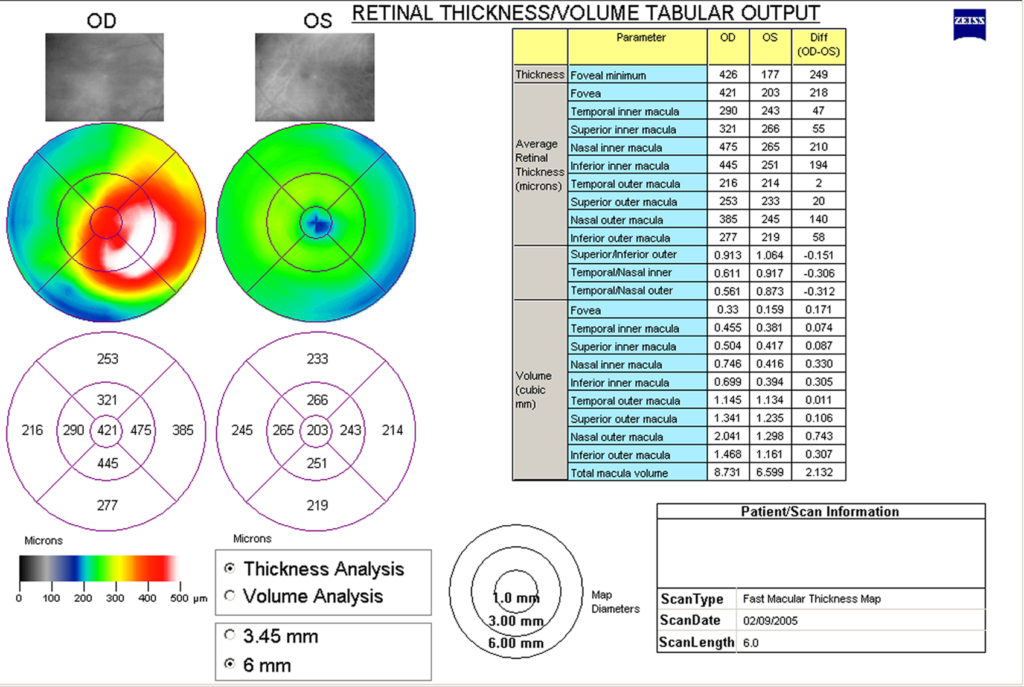
OCT was carried out at the 10 week review. Fast macular thickness map revealed a raised ovoid area of neuro-sensory detachment extending from the disc to just beyond the nasal fovea along temporal arcade and disc, corresponding to expected site of photo-toxic damage from temporal clear corneal approach used for surgery. The most affected sector was nasal inner macula; mean retinal thickness was 475µ compared to 265µ on the left (Fig. 2C). A 7mm OCT image scan revealed a dome shaped detachment of neurosensory retina. There was no evidence of thickening of the neurosensory retina, or any cystoid spaces (Figure 2D).
Discussion:
Phototoxic retinopathy was originally described after extracapsular (ECCE) surgery(4), and two subsequent prospective studies found 7% and 28% of cases had evidence of phototoxic injury(1, 6). Although the risk of phototoxicity has decreased with phacoemulsification surgery of short duration, at least one study has demonstrated that phototoxicity may occur (3).
The OCT findings of the above cases suggest a pattern of changes in photic injury. The main finding was of retinal thickening which consisted of two components; development of neuro-sensory detachment from underlying RPE in the affected retina, with lesser and varying degree of neural retinal thickening and cystoid change. The area of thickening and elevation coincided with subtle RPE pigmentary changes on fundus examination. The changes corresponded to area of maximum intensity by the coaxial beam of microscope on the retina and were not centred on the fovea.
The pathophysiological basis of phototoxic retinopathy has been the subject of considerable study(7, 8). Animal and human histopathologic studies have suggested that the site of damage may be the RPE(9, 10), although this is uncertain(10). The presence of neurosensory detachment with collection of fluid between neural and RPE layer in our study may suggest that RPE decompensation due to photoxicity may be the prime site of pathology. However, it remains debatable whether the previously described histologic changes may be secondary to functional damage in an adjacent layer. Huang et al(11) described OCT findings in foveal phototoxic injury induced by solar, welding and laser exposure, while Bechmann et al(12) and Kaushik et al (13) have described OCT findings in solar retinopathy. In these cases, the lesions were much more focal due to the nature of the injury, but correspond with our findings in that the site of OCT lesion was outer retina/inner RPE. Neuro sensory detachment, as in our cases, was absent in such focal injuries. Huang et al also noted the limitations of fluorescein/ICG angiographic analysis in this regard (11).
OCT is an imaging modality allowing high resolution cross sectional imaging of the retina in vivo. It provides accurate retinal thickness measurement, and limited representation of the histologic structures of the retina (14, 15). In this regard, the changes outlined above are likely to represent sub-neuroretinal fluid collection secondary to RPE functional compromise, or oedema/tissue destruction of outer neuroretinal layers. However, known limitations of current OCT technology in defining histological layers do not allow concrete conclusions to be drawn (14).
References
- Byrnes GA, Antoszyk AN, Mazur DO, Kao TC, Miller SA. Photic maculopathy after extracapsular cataract surgery. A prospective study.
- Khwarg SG, Linstone FA, Daniels SA, Isenberg SJ, Hanscom TA, Geoghegan M, et al. Incidence, risk factors, and morphology in operating microscope light retinopathy. Am J Ophthalmol. 1987;103(3 Pt 1):255-63.
- Kleinmann G, Hoffman P, Schechtman E, Pollack A. Microscope-induced retinal phototoxicity in cataract surgery of short duration. Ophthalmology. 2002;109(2):334-8.
- McDonald HR, Irvine AR. Light-induced maculopathy from the operating microscope in extracapsular cataract extraction and intraocular lens implantation.
- Gomolin JE, Koenekoop RK. Presumed photic retinopathy after cataract surgery: an angiographic study. Can J Ophthalmol. 1993;28(5):221-4.
- Byrnes GA, Chang B, Loose I, Miller SA, Benson WE. Prospective incidence of photic maculopathy after cataract surgery. Am J Ophthalmol. 1995;119(2):231-2.
- Michels M, Sternberg P, Jr. Operating microscope-induced retinal phototoxicity: pathophysiology, clinical manifestations and prevention. Surv Ophthalmol. 1990;34(4):237-52.
- Verma L, Venkatesh P, Tewari HK. Phototoxic retinopathy. Ophthalmol Clin North Am. 2001;14(4):601-9.
- Green WR, Robertson DM. Pathologic findings of photic retinopathy in the human eye. Am J Ophthalmol. 1991;112(5):520-7.
- Ham WT, Jr., Ruffolo JJ, Jr., Mueller HA, Clarke AM, Moon ME. Histologic analysis of photochemical lesions produced in rhesus retina by short-wave-length light. Invest Ophthalmol Vis Sci. 1978;17(10):1029-35.
- Huang SJ, Gross NE, Costa DL, Yannuzzi LA. Optical coherence tomography findings in photic maculopathy. Retina. 2003;23(6):863-6.
- Bechmann M, Ehrt O, Thiel MJ, Kristin N, Ulbig MW, Kampik A. Optical coherence tomography findings in early solar retinopathy. Br J Ophthalmol. 2000;84(5):547-8.
- Kaushik S, Gupta V, Gupta A. Optical coherence tomography findings in solar retinopathy. Ophthalmic Surg Lasers Imaging. 2004;35(1):52-5.
- Chauhan DS, Marshall J. The interpretation of optical coherence tomography images of the retina. Invest Ophthalmol Vis Sci. 1999;40(10):2332-42.
- Toth CA, Narayan DG, Boppart SA, Hee MR, Fujimoto JG, Birngruber R, et al. A comparison of retinal morphology viewed by optical coherence tomography and by light microscopy. Arch Ophthalmol. 1997;115(11):1425-8.
