Brendan Leng Yong Ji, Diya Baker
Background
Dome shaped maculopathy (DSM) is a convex anterior protrusion of the macula into the vitreous cavity. DSM is commonly associated with high myopia and posterior staphyloma but can occur in emmetropia. DSM is thought to occur in 12% of highly myopic eyes but the exact population prevalence is currently unknown (1). The major complication of DSM is serous retinal detachment, likely secondary to subretinal fluid (SRF) accumulation (2). DSM is an unusual, poorly understood yet relatively common disease that most Ophthalmologists will encounter in their practice.
Case Presentation
A 55-year-old lady with a history of high myopia and previous cataract surgery is referred by optometry after an incidental finding of SRF in her left eye on ocular coherence tomography (OCT). She has a past medical history of asthma but is otherwise fit and well. She was asymptomatic at the time of contact with no visual or systemic symptoms. Socially she works as a teacher, is mobile and independent with no major life stressors.
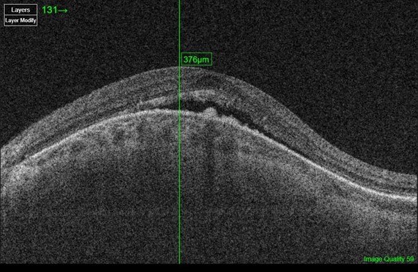
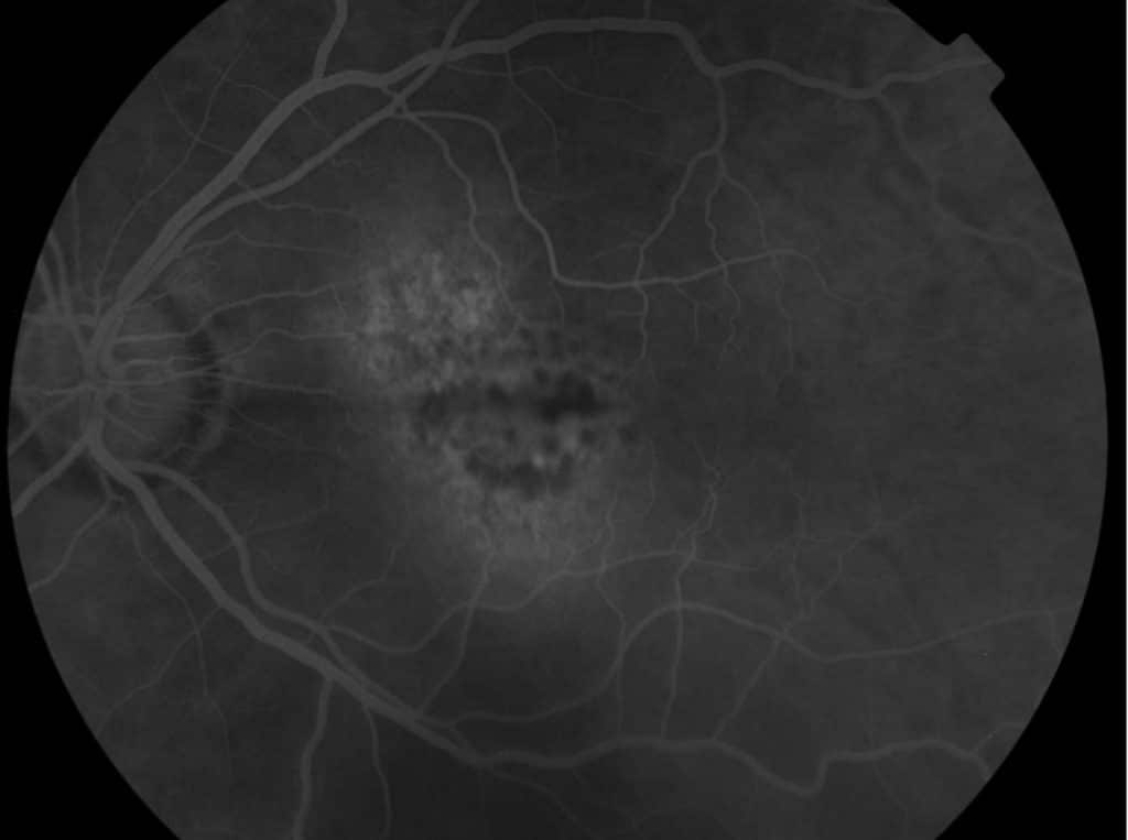
On examination, her best corrected visual acuity (BCVA) is 6/6 OD and 6/7.5 OS. Anterior segment examination was normal. Dilated fundus examination showed myopic discs bilaterally with dome shaped maculopathy in the left eye. Testing with Amsler grid showed no signs of metamorphopsia bilaterally. OCT reinforced diagnosis of DSM with characteristic anterior convexity of macula with associated SRF (figure 1). Further investigation with fluorescein angiography (FFA) identified active areas of leakage in the macula (figure 2) and hence a decision to treat with intravitreal anti-vascular endothelial growth factor (anti-VEGF) injections. She will be actively monitored and followed up in the medical retina clinic to evaluate her response to treatment.


The visual prognosis of DSM is relatively positive, with SRF complications being the main cause of visual deterioration (3). As such, the mainstay of treatment is aimed at reducing SRF, but initiation of treatment is largely to the clinician’s discretion due to lack of accepted guidelines. Treatment options utilized include intravitreal Anti-VEGF and photodynamic therapy (PDT). However, clinical efficacy of available treatment is equivocal (3-4). The aim of decreasing SRF is to minimize the risk of serous retinal detachment. There is conflicting evidence on whether the presence of SRF is associated with decreased BCVA (5-6).
Learning points
DSM is a relatively common condition that requires appropriate recognition and follow up. OCT and FFA are vital ancillary investigations in the workup of DSM. Not all patients are candidates for intervention, but treatment choices could include anti-VEGF injections and PDT.
References
- Gaucher D, Erginay A, Lecleire-Collet A, et al. Dome-shaped macula in eyes with myopic posterior staphyloma. Am J Ophthalmol. 2008;145(5):909-914.
- Lorenzo D, Arias L, Choudhry N, et al. Dome-shaped macula in myopic eyes: twelve-month follow-up. Retina. 2017;37(4):680-686.
- Lorenzo D, Arias L, Choudhry N, Millan E, Flores I, Rubio M et al. DOME-SHAPED MACULA IN MYOPIC EYES. Retina. 2017;37(4):680-686. Giacomelli G, Mencucci R, Sodi A, et al. Aflibercept in Serous Foveal Detachment in Dome-Shaped Macula: Short-Term Results in a Retrospective Study. Ophthalmic Surg Lasers Imaging Retina. 2017;48(10):822-828.
- Arapi I, Neri P, Mariotti C, et al. Considering photodynamic therapy as a therapeutic modality in selected cases of dome-shaped macula complicated by foveal serous retinal detachment. Ophthalmic Surg Lasers Imaging Retina. 2015;46(2):217-223.
- Caillaux V, Gaucher D, Gualino V, Massin P, Tadayoni R, Gaudric A. Morphologic characterization of dome-shaped macula in myopic eyes with serous macular detachment. Am J Ophthalmol.
